Optomap Ultrawide Scan - Optos California
Experience Advanced Retina Technology at Eye Academy with Optomap Scans.
Optomap is the latest imaging technology available at Eye Academy Windsor, which, when combined with Spectralis OCT examination, allow a comprehensive view of the retina, the inner lining of the eye that is essential for vision. This technology allows for a quick, painless, and non-invasive examination that can detect potential disease before it becomes a serious sight-threatening condition.
At Eye Academy in Windsor, we are proud to be the only opticians in the area to offer Optomap technology, via the Optos California to our patients. By including Optomap in your eye examination, you can ensure that your retina is healthy and that any potential issues are caught early.
What is an Optomap Scan?
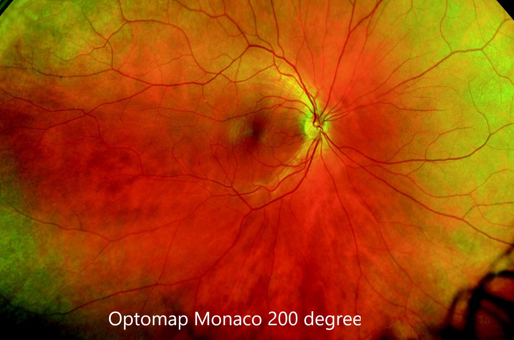
An Optomap Scan is a state-of-the-art technology that allows our clinicians to take a 270-degree ultra-widefield, highly detailed and comprehensive image of the retina, the light-sensitive layer at the back of the eye. This technology is designed to help detect potential issues with the retina such as retinal tears and detachments and monitor other eye conditions such as diabetic retinopathy and retinal naevi.
What Happens During an Optomap Scan?
During an Optomap scan, you will be asked to look directly at a target in the device that takes an ultra-widefield, high-definition image of your retina. The process is quick and painless, taking less than a minute to complete. The ultra-widefield Optomap image can then be analysed by our expert clinicians who have extensive experience utilising these scans to diagnose and detect any potential issues.
What Conditions Does an Optomap Scan Help to Diagnose?
Optomap scans can help diagnose a wide range of conditions that affect the retina and your overall general health. As well as monitoring commonly found eye condition such as macula degeneration, myopic retinal degeneration, retinal atrophy and retinal schisis, the role of Optomap really lies in its ability to identify serious pathology that can affect both your eyes and general health.
Sight-threatening diseases that can be diagnosed through an Optomap exam includes the following:
Retinal tears
Retinal Detachments
Diabetic Retinopathy
Retinal bleeds
Life-threatening pathology that can be identified through an Optomap exam includes the following:
Eye Cancer
Eye Cancers are difficult to identify without a comprehensive peripheral examination that includes an ultrawide field, and dilated 200-degree retinal examination.
Find out more about Optomap scans and Melanoma here.
Cardiovascular Disease
The retina is the only place in the body that allows non-invasive examination of the body’s arteries and veins in situ. As your eye health is connected to your heart health, retinal vasculature analysis can provide an indicator of cardiovascular health. Analysis and comparison of retinal arteries and veins are now used in screening as a predictive factor in determining cardiovascular health.
Stroke
Optomap UltraWideF examinations allow detailed examination of retinal vessels, including blockages. Blockages in retinal vasculature can lead to sight-threatening conditions such as retinal vein and artery occlusions (a stroke in the eye that is a medical emergency) and can be a predictive factor for stroke.
What Is the Difference Between Optomap Scans and Traditional Digital Retinal Photography?
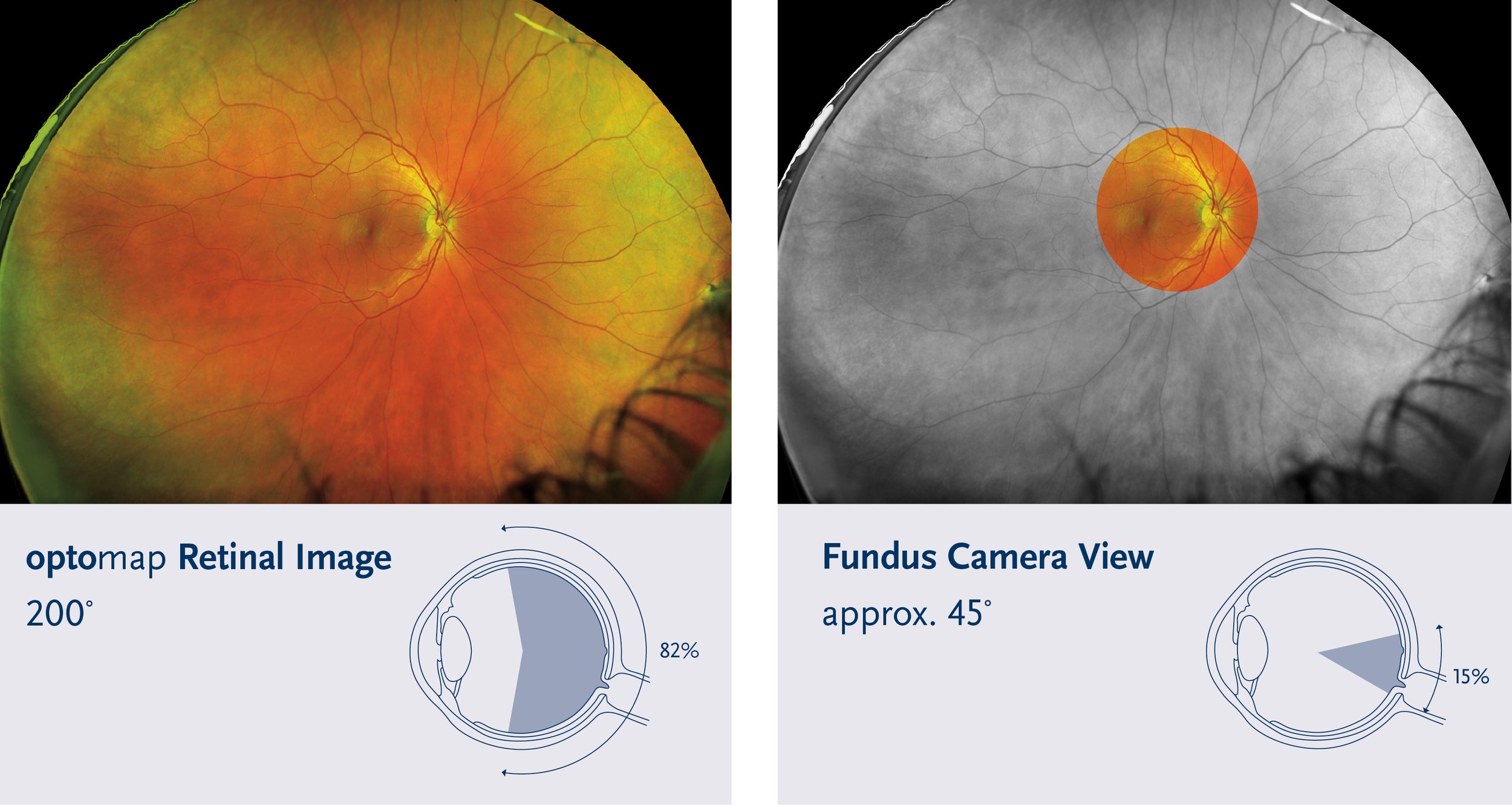
Fundus photography is now considered legacy technology versus Optomap which has become the gold standard for peripheral retinal examination. Both Optomap scans and digital retinal photography provide an image of the retina, but the Optomap scan gives a much wider and more detailed retinal view versus the limited view generated with fundus photography. An Optomap ultra-widefield digital retinal imaging system captures more than 80% of your retina in one panoramic image. Traditional methods typically reveal only 10-15% of your retina at one time.
Optomap view vs. Traditional Fundus Photography
An Optomap scan can be compared to a panoramic photo of a cityscape, taken with one of the best cameras in the world, where the entire view of the retina is captured in one ultra-high-definition image. Digital retinal photography, on the other hand, can be compared to a standard definition, zoomed-in photograph of a specific building within the cityscape.
What Is the Difference Between Optomap and OCT?
An Optomap scan can be compared to a full-body scan in a hospital, providing a broad overview of the retina. On the other hand, OCT (Optical Coherence Tomography) is like an x-ray, providing a cross-sectional detailed view of the retina. Both are vital in diagnosing and detecting any issues.
Do You Really Need Both an Optomap Scan and An OCT Scan?
Yes! Having both an Optomap scan and an OCT (Optical Coherence Tomography) scan provides a more comprehensive understanding of the health of the retina.
An Optomap scan provides a wide-angle, high-resolution image of the retina, allowing our optometrists to see the entire retina in one image. This can help detect any issues that may not be visible with traditional methods, such as small changes in the retina or subtle signs of disease.
On the other hand, an OCT scan provides a cross-sectional image of the retina, which is useful for seeing the different layers of the retina in detail. This can help detect issues that may not be visible on an Optomap scan, such as thinning of the retina or fluid build-up.
Together, the Optomap and OCT scans provide a more complete picture of the retina. The Optomap scan gives a broader overview of the retina, and the OCT scan provides detailed information about specific areas. Having both scans can help detect issues early on, allowing for timely treatment and better outcomes.
Both the Optomap scan and the OCT scan have unique features and provide different information, together they help to give a complete picture of the retina, which can help to detect and diagnose issues early on and improve the outcome of treatment and management.
Who Should Have an Optomap Scan?
Everyone! An Optomap scan is a great way for people to proactively monitor the health of their eyes and catch any issues early on.
More specifically, anyone who is concerned about the health of their eyes, or falls into the following groups, would benefit from an Optos California Optomap scan at Eye Academy Windsor.
Short-sightedness (myopia), including those who have had laser eye surgery- the shorter-sighted you are, the more at risk you are of retinal pathology hence an Optomapscan is a must for all short-sighted individuals.
All diabetic patients
All patients who have been diagnosed with hypertension or cardiovascular disease.
New or long-standing floaters
New or long-standing flashing lights (photopsia)
Those with a history of retinal tear of detachments
Those with a family history of retinal tear of detachments
Those with a history of retinal freckles/moles, eye tumours or cancer
Those with a family history of eye cancers
Should My Children Have an Optomap Too?
Yes! Many vision problems begin at an early age, so it’s important for children to receive proper eye care from the time they are infants. Early detection and treatment are essential to preventing conditions that could potentially cause problems or vision loss. The state-of-the-art Optos ultra-widefield (UWF™) imaging technology is allowing our eye examinations to be quicker and more comfortable for children.
Optomap was designed by Douglas Anderson, who was driven by his son Leif's experience. Leif's loss of vision in one eye due to a late-detected retinal detachment spurred Douglas to create a better solution. Routine eye exams, especially for young children, can be uncomfortable, hindering a comprehensive examination. Douglas aimed to capture as much of the retina as possible in a non-invasive manner, leading to the birth of Optomap.
Unlike traditional fundus imaging, which can be complex and challenging for children, an Optomap image offers a panoramic view, capturing even the periphery with a single shot. This streamlines the imaging process, making it quicker and more comfortable. Backed by multiple clinical studies, Optomap has emerged as a pivotal tool in screening and managing pediatric patients.
What Makes the Optomap California Different?
Just like OCT scans, Optomap scans are not all the same. You can get different levels of Optomap scans that differ in quality and ability. At Eye Academy, we have invested in one of the best, most advanced Optomap Scanners called the Optos California.
Optomap scans captured via the Optos California are 200-degree, ultrawide, ultra-high-definition image, and scan quality and field of view is far superior to other Optomap images. Additionally, it is equipped with Optos Advanced software that allows for a more detailed analysis of the retina, further increasing its diagnostic capabilities. With its cutting-edge technology, the Optomap California is the most advanced option available for monitoring and diagnosing retina-related issues.
Other Opticians have Optomap- why is Eye Academy different?
As with all aspects of medicine, quality imaging is important, but it is the clinician’s image interpretation and analysis of the images that allow for accurate eye disease detection and monitoring.
Here at Eye Academy Windsor, all our clinicians are advanced, specialist optometrists that undergone specific further training. Most of our clinicians have, or continue to hold specialist positions in eye hospitals, where they detect and manage complex eye disease as part of a wider multi-disciplinary team. Eye Academy Windsor does not employ general optometrists and all clinical staff are specialists registered on the GOC specialist register.
This skill level allows Eye Academy Windsor to offer a clinical service comparable to that of a hospital eye service, with specialist assessments for differing eye conditions.
A combination of expert clinicians and the latest diagnostics equipment allows us to be experts in our fields.


